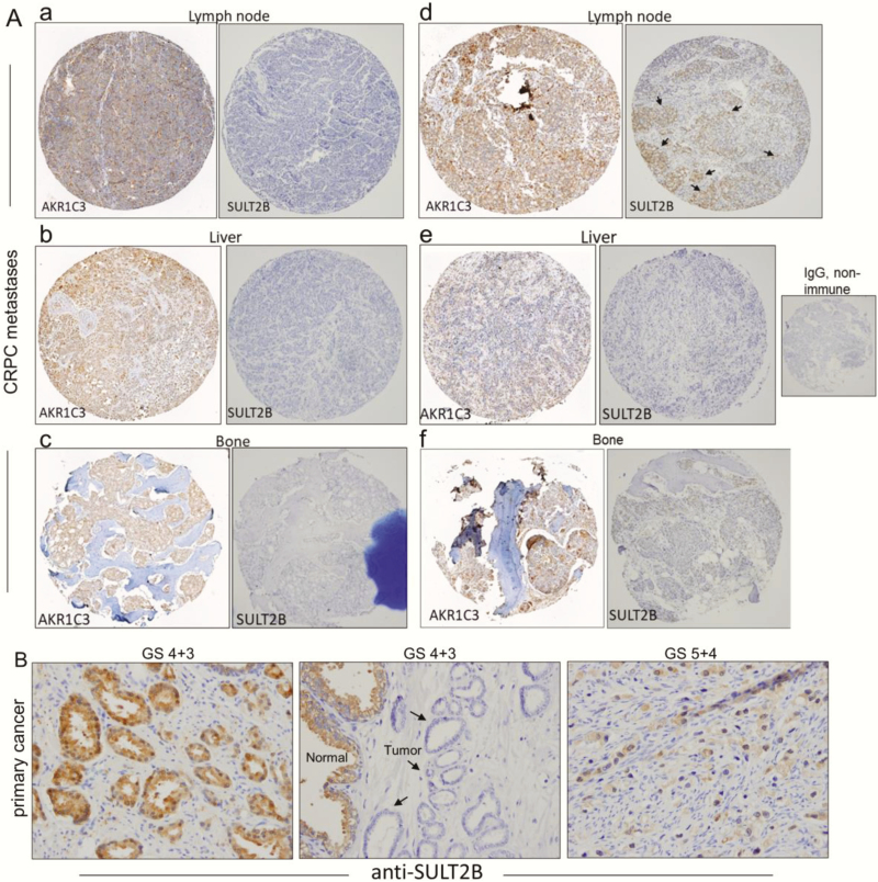
Figure 7.
Loss of SULT2B in AKR1C3-positive clinical CRPC metastases and variable SULT2B status of primary prostate cancer. (A) IHC of lymph node, liver and bone metastases of CRPC. Each panel with 2 tissue cores from the same specimen shows staining for AKR1C3 (left) and SULT2B (right). For panels (a–c and e,f), specimens are SULT2B-negative, while AKR1C3 expression is relatively strong. In panel d, the AKR1C3-positive CRPC showed limited SULT2B staining (marked by arrows). The non-immune IgG negative control is shown. (B) SULT2B in primary prostate tumor. Middle panel: SULT2B-negative tumor epithelia (shown by arrows) for a GS 7 specimen. Right panel: poorly differentiated GS 9 tumor showing SULT2B-positive status. Left panel: A second GS 7 specimen showing strong SULT2B staining in tumor acini.

