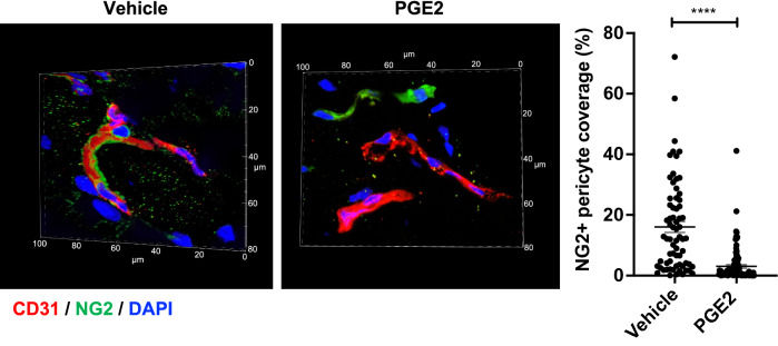
Figure 3.
Dermal capillary vessels lose pericyte coverage upon local PGE2 injection. Mice were injected with PGE2 (200 ng) or vehicle (DMSO) into dorsal skin on Day 0 and Day 1, then skin samples were collected and tissue sections were immunostained for NG2 (green), CD31 (red) and DAPI (blue). A representative micrograph of microvessels is shown for each condition. The pictures were taken using a 60 × objective lens. The depth of confocal scanning was 4.35 μm; therefore, winding microvessels are seen in fragments in these pictures. The right picture panel shows NG2+ pericytes that are dissociated from the CD31+ endothelium in the PGE2-treated skin. Blood vessel coverage by NG2+ pericytes is presented in a graph with an arbitrary unit. The analysis was conducted in total of > 68 micrographs of different areas in 5 skin samples for each condition. Each dot represents one micrograph. Student t-test, ****p < 0.0001.

