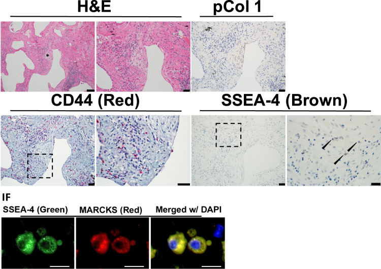
Figure 6.
Cells expressing CD44, SSEA-4 and MARCKS reside in the cellular perimeter region of the fibroblastic focus. An Idiopathic Pulmonary Fibrosis (IPF) specimen was serially sectioned at 4 μm and processed for histology, immunohistochemistry (IHC) and immunofluorescence (IF). IHC: Representative images for Hematoxylin and Eosin (H&E) staining (scale bar represents 50 μm left and 20 μm right) with an asterisk labeling a fibroblastic focus; Immunostaining for anti-procollagen type I (brown, scale bar 20 μm); anti-CD44 (red, scale bar 20 μm, dashed outline box, scale bar 20 μm); anti-SSEA4 (brown, scale bar 20 μm, dashed outline box, scale bar 10 μm). Lower panel: Immunostaining anti-SSEA-4 (green), MARCKS (red), DAPI (blue, scale bar 20 μm). A small apoptotic body is noted adjacent to the cell on the right. Immunofluorescence images obtained at the perimeter of the fibroblastic focus.

