FIGURE 1.

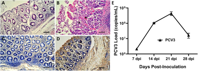
Histopathological lesions and immunohistochemical staining of small intestines and porcine circovirus type 3 (PCV3) loads in sera of PCV3- and sham-inoculated piglets. (A) Normal morphology of a small intestine section from a sham-inoculated piglet. (B) Small intestine lesions from a PCV3-inoculated piglet showed mucosal epithelial cell necrosis and some necrosis of lymphocytes, infiltration of abundant eosinophils and lymphocytes, and small numbers of plasma cells (arrowheads). (C) No staining was observed in the small intestine section from a sham-inoculated piglet. (D) Several PCV3 antigen-positive cells (arrowhead) were also observed in the small intestine of a PCV3-inoculated piglet, which were stained brown. Bars, 80 μm. (E) Viral loads in sera were quantitatively assayed by real-time PCR in PCV3-inoculated piglets. The values presented are the means of the results from the four PCV3-inoculated piglets at 7, 14, 21, and 28 dpi. Error bars indicate standard deviations.
