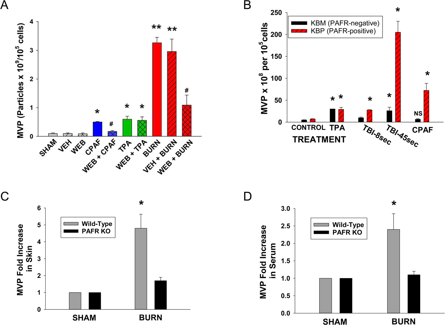
Figure 3. Thermal burn injury of epithelial cells induces MVP in a PAFR-dependent manner.

(A) HaCaT cells were treated with no treatment (sham), control (0.1% ethanol) vehicle, or preincubated with 10 μM of the PAFR antagonist WEB 2086, or 100 nM CPAF, 100 nM TPA, thermal burn injury (45 sec at 90 °C), or WEB 2086 followed 1 h later by the above agents. Two hours post-treatment, supernatants were removed and MVP quantified. (B) KBM and KBP cells were treated with control vehicle, 100 nM CPAF, 100 nM TPA, or thermal burn injury of 8 or 45 sec. Four hours later MVP were isolated from supernatants. The data are Mean ± SE MVP levels from at least four separate experiments. (C, D) Wild-type or PAFR KO mice (Ptafr−/−) mice were subjected to a thermal burn injury to approximately 15–20% body surface area, and 2 h later (C) three 5 mm punch biopsies or (D) serum was tested for MVP. The data are Mean ± SE MVP levels normalized to SHAM-treated values from at least 6–8 mice in each group. Statistically significant (*: P<0.05; **: P < 0.01) changes in levels of MVP from control (SHAM-treated) values. # Denotes statistically significant (P<0.05) differences in vehicle vs WEB 2086 treatment. N.S., not statistically significant.
