
Figure 3.
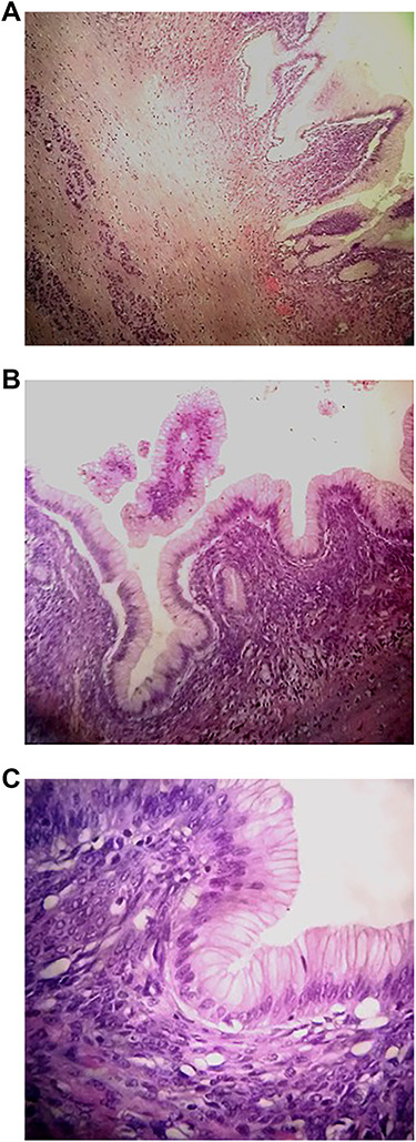
Histological analysis. A: Pancreatic cystic wall (Hematoxylin and eosin stain (HE); ×40 magnification). B: Pancreatic cystic wall lined by foveolar gastric type surrounded by dense ovarian type stroma (HE; ×100). C: Details of epithelium with low grade dysplasia (HE; ×400).