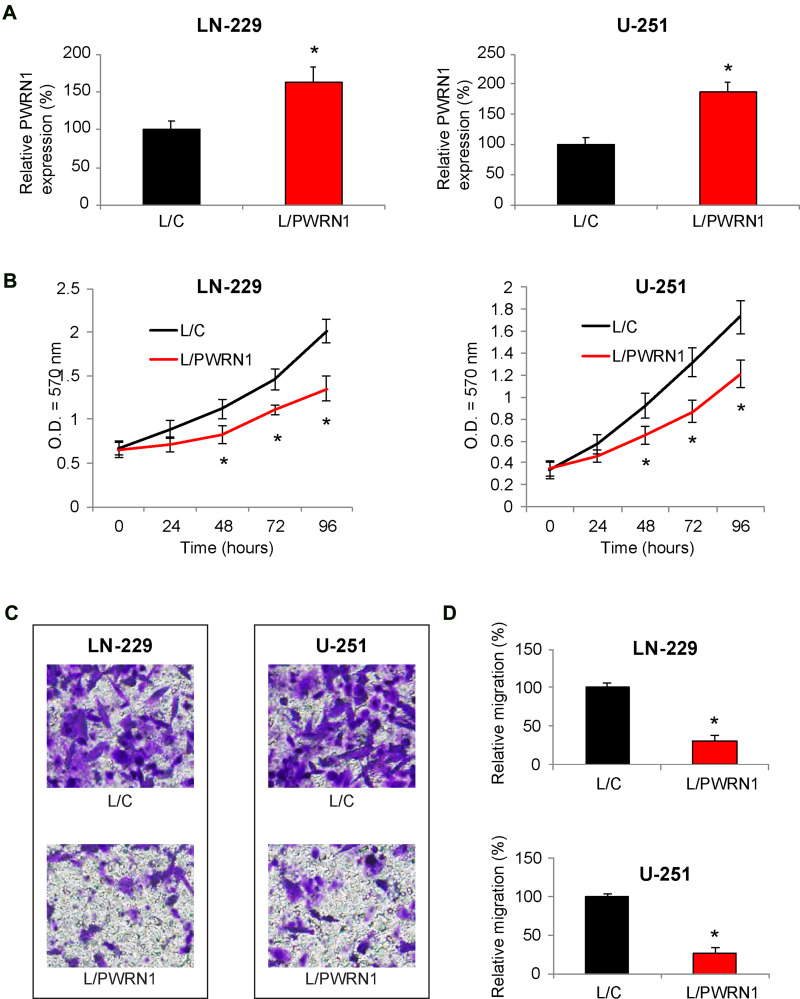
Figure 2.
Inhibitory effects of PWRN1 overexpression on GBM cell proliferation and migration in vitro. (A) LN-229 and U-251 cells were infected with L/C or L/PWRN1. Post infection, cells were passaged 5~8 times, followed by qRT-PCR to assess theirPWRN1expressions (* P < 0.05). (B) InfectedLN-229 and U-251 cells were assessed by a CCK-8 assay for 96 h, to compare in vitro proliferation between L/C- and L/PWRN1- infected GBM cells (* P < 0.05). (C) InfectedLN-229 and U-251 cells were assessed by a 24-well transwell assay. After 24 h, LN-229 and U-251 cells migrated onto the bottoms of wells were stained. Representative images were shown for L/C- and L/PWRN1- infected GBM cells. (D) Formigrating LN-229 and U-251 cells shown in (C), relative migration was quantitatively measured and compared between L/C- and L/PWRN1- infected GBM cells (* P < 0.05).

