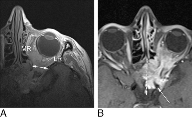
Fig 2.

A, MC-MR imaging T1-weighted axial image demonstrates a sphenoid wing meningioma, engulfing the optic nerve as well as the medial (MR) and lateral rectus (LR) muscles. MC-MR imaging demonstrates widening of the superior orbital fissure (double-headed arrow), but beyond the orbital apex, the signal-to-noise ratio is low. B, Head coil T1-weighted fat-saturation postcontrast axial image demonstrates the lesion surrounding the left internal carotid artery (white arrow), infiltrating the left cavernous sinus and extending past the midline (dotted white line) in the intercavernous sinus.
