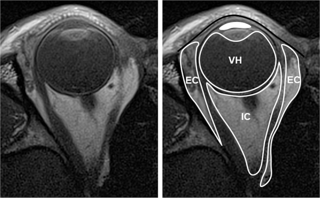
Fig 6.

Compartmental anatomy shown by MC-MR imaging T1-weighted axial image, original on the left and annotated on the right. The solid black line indicates the orbital septum, defined by native high signal of the tarsal plate; EC, extraconal space, external to the extraocular muscles; IC, intraconal space, inside the extraocular muscles; VH, vitreous humor, behind the lens; solid white fill, aqueous humor, anterior to the lens and ciliary muscles.
