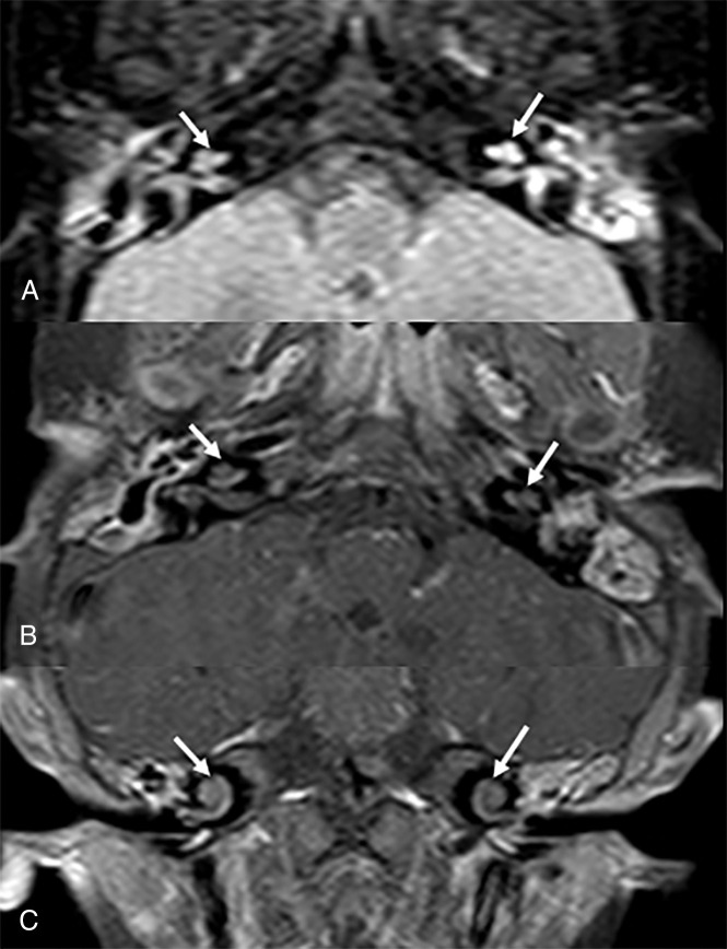
Fig 3.
Seven-month-old girl with history of meningitis from N meningitidis. (A) Magnified axial FLAIR, (B) axial T1-weighted+C, and (C) coronal T1-weighted+C images demonstrate abnormal hyperintense FLAIR signal and enhancement of both inner ears. There is an increased enhancement on the mastoids bilaterally. The patient developed bilateral SNHL on follow-up.

