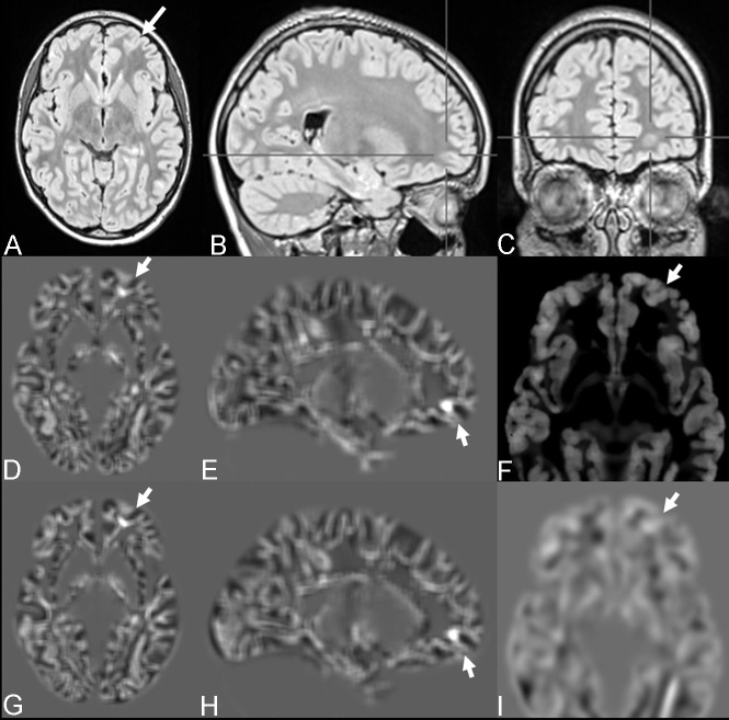
Fig 1.
FCD with the transmantle sign of the left medial fronto-orbital gyrus (bottom-of-sulcus dysplasia) in a 12-year-old girl with seizures (patient 1). The lesion was overlooked on visual inspection (A–C) but was highlighted with MPRAGE-based (D and E) and MP2RAGE-based (G–H) junction images. MP2RAGE-based thickness (F) and extension (I) maps were considered to have negative findings.

