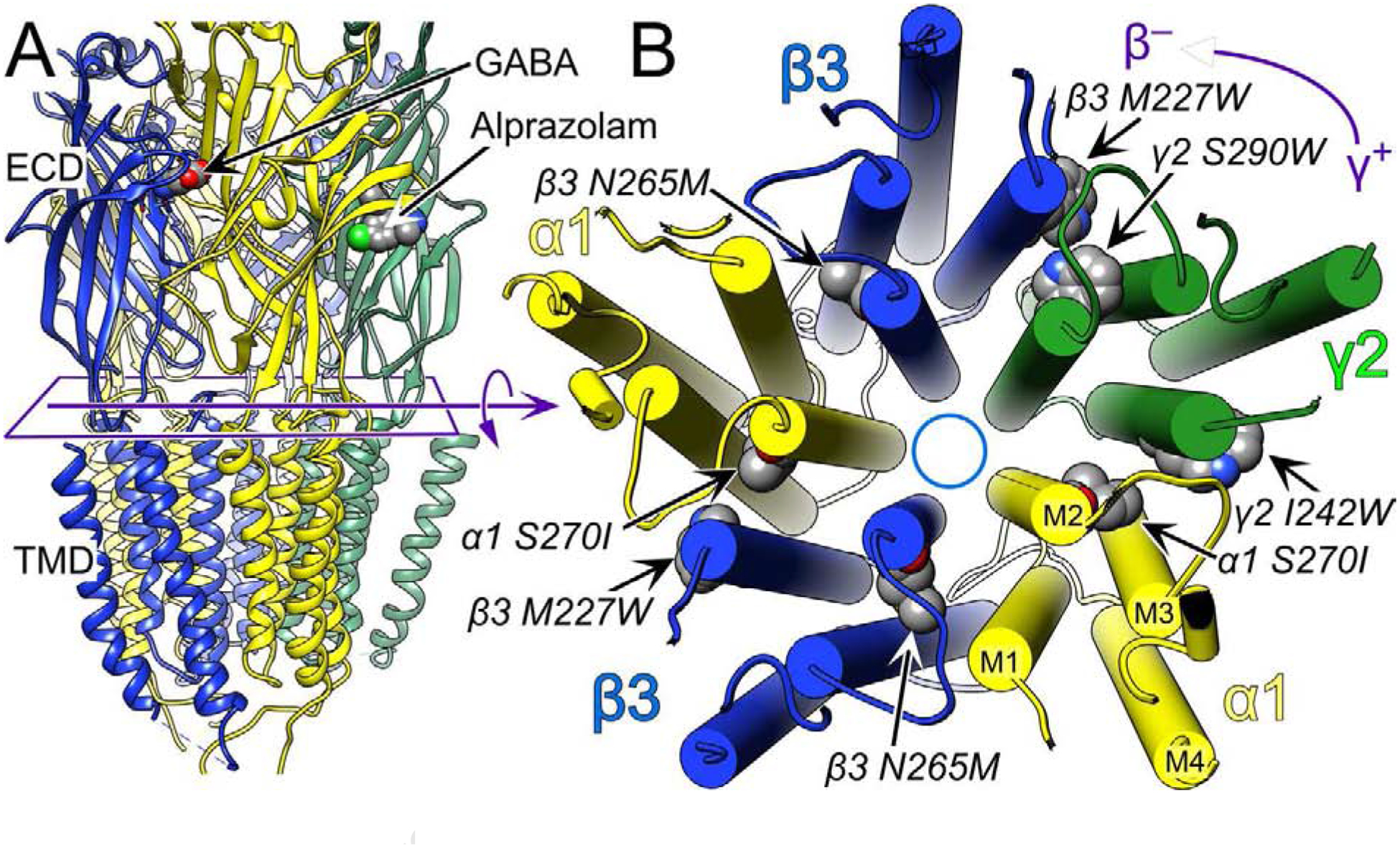
Figure 1. The structure of the human full length α1β3γ2L GABAAR showing the main amino acid residues mentioned in this manuscript.

Panel A shows a side view of the extracellular domain (ECD) and transmembrane domain (TMD) depicted in ribbon mode. The intracellular domain is unstructured and therefore not shown. Panel B shows a cross section of the TMD viewed from the extracellular side with cylindrical helices. The subunits are labeled and color coded as indicated. The convention is to refer to the subunits in counter clockwise order with the plus and minus side of the subunit interface defined as indicated in the top right corner, which defines the γ+/β− subunit interface. In the ECD, GABA binds to the two β+/α− interfaces and alprazolam, a benzodiazepine, binds in the single α+/γ− interface. The structure shown is from the Protein Data Base, 6HUO.pdb, which is the α1β3γ2L GABAAR with both GABA and Alprazolam bound [7]. The figure was created using UCSF Chimera, developed by the Resource for Biocomputing, Visualization, and Informatics at the University of California, San Francisco, with support from NIH P41-GM103311 [39].
