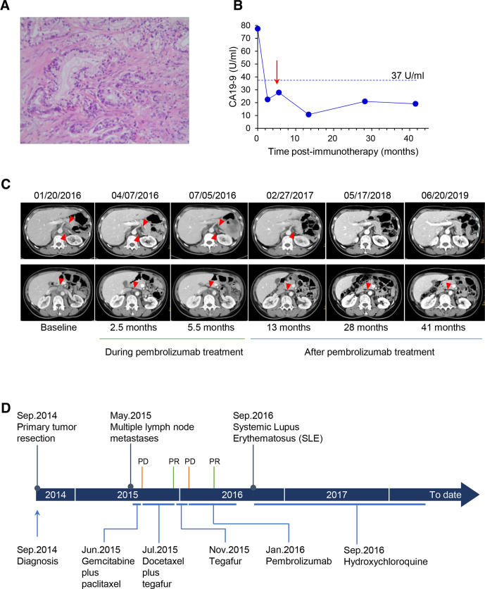
Figure 1.
Clinical course of the patient. (A) Histological characteristic of the moderately differentiated adenocarcinoma of the pancreas (hematoxylin-eosin stain; original magnification×400). (B) Serum CA19-9 levels during and after treatment with pembrolizumab (red arrow indicates the last dose of pembrolizumab). (C) Representative contrast-enhanced CT images of the abdomen in venous phase, showing patient’s recurrent lymph node lesions during and after treatment with pembrolizumab. A 2.5-month follow-up scan demonstrated partial response of the disease. (the top row: retroperitoneal lymph nodes; the bottom row: abdominal para-aortic lymph node; lesions are marked by red triangle). (D) Time line of clinical events. PD, progressive disease; PR, partial response.

