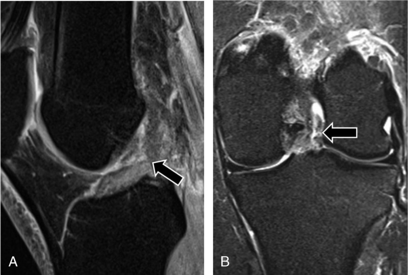
FIGURE 3.

MRI of the left knee of a 41-year-old woman with knee injury 1 week earlier. Sagittal intermediate-weighted turbo spin echo image with fat suppression (A) and coronal short tau inversion recovery image (B) show a full-thickness tear of the midsubstance of the ACL (arrows), which was confirmed by arthroscopic surgery. The DCNN and all 3 radiologists correctly diagnosed the full-thickness ACL tear.
