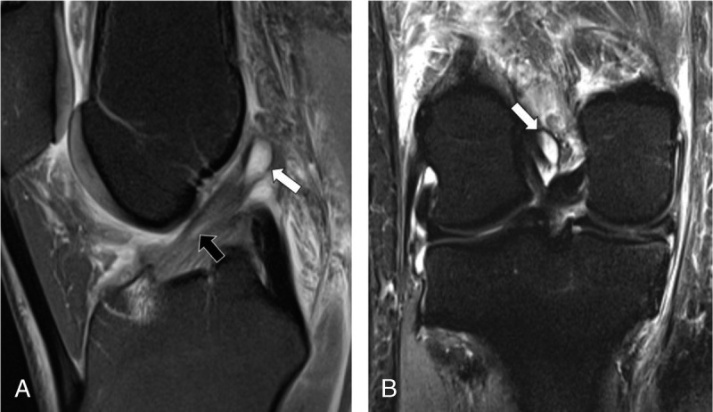
FIGURE 5.

MRI of the right knee of a 43-year-old woman with knee injury 7 days earlier. Sagittal intermediate-weighted turbo spin echo image with fat suppression (A) and coronal short tau inversion recovery image (B) show diffuse and focal (black arrow) signal hyperintensity of the anterior cruciate ligament (ACL) indicative of mucoid degeneration and an intraligamentous ganglion cyst (white arrows) with otherwise continuous fibers in normal oblique orientation. Arthroscopic surgery confirmed mucoid degeneration of the ACL without fiber discontinuity. All 3 radiologists correctly diagnosed an intact ACL, whereas the DCNN erroneously classified the ACL as torn, representing a false-positive case.
