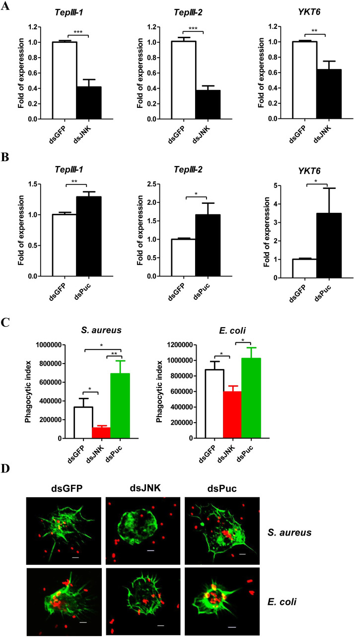
Fig 4. JNK pathway mediates hemocytes phagocytosis.
Effect of JNK (A) and Puc (B) silence on the mRNA levels of phagocytosis related genes: TepIII-1, TepIII-2 and YKT6 of the pea aphids. The expressions of TepIII-1, TepIII-2 and YKT6 were normalized with rpl7 of the pea aphids. (C) Ex vivo phagocytosis assay using S. aureus and E. coli AlexaFluor 594 BioParticle (Invitrogen) after knockdown of JNK and Puc. The hemocytes from 20 pea aphids per group were used to perform each experiment. In (A-C), the values shown are the mean (±SEM) of three independent experiments and the statistical differences between the compared groups were denoted with asterisks. P-values were determined by Student’s t test. *P<0.05; **P<0.01; ***P<0.001. (D) The photographs of ex vivo phagocytosis S. aureus and E. coli AlexaFluo 594 BioParticles (Invitrogen) by the hemocytes with the F-actin stained by SF-488 Phalloidin (1/200 diluted, Solarbio) after knockdown of JNK and Puc. The red dots were S. aureus and E. coli, and the green parts were the hemocytes with the F-actin stained. Scale bar: 5 μm.

