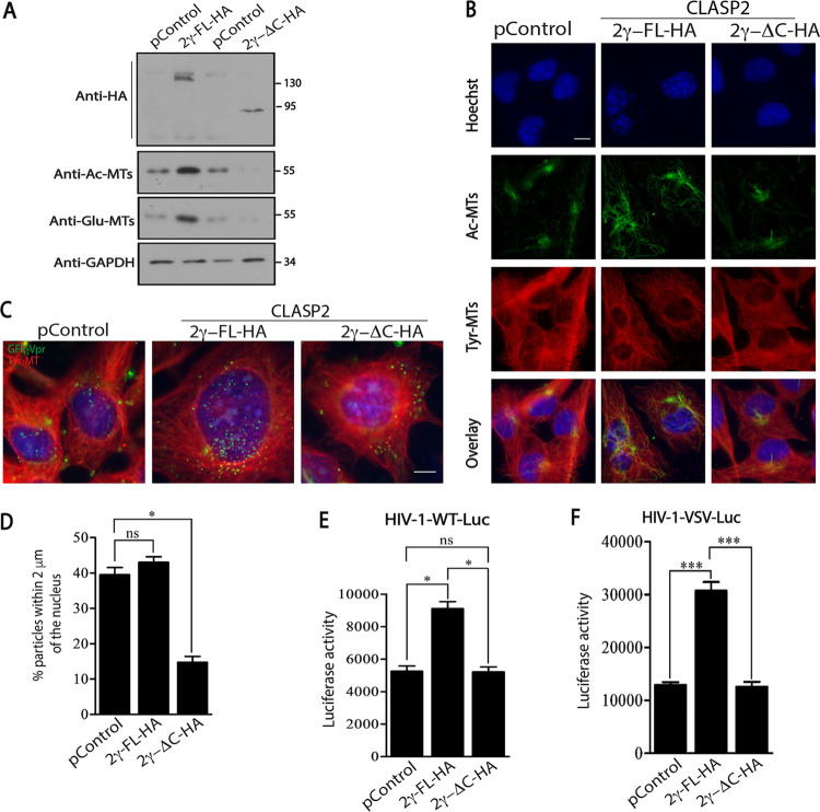
FIG 4.
C terminus of CLASP2γ induces stable MTs to promote early HIV-1 infection. (A and B) The levels of tyrosinated tubulin (Tyr-MTs), acetylated tubulin (Ac-MTs), or detyrosinated tubulin (also known as Glu-MTs) detected in CHME3 cells stably expressing empty vector control (pControl), 2γ-FL-HA, or 2γ-ΔC-HA by WB analysis (A) or IF assay (B). Molecular-weight markers (in kDa) are shown to the right of WBs. Representative (n = 3) staining images for Tyr-MTs, Ac-MTs, or nuclei (Hoechst stain) are shown. Scale bar = 10 μm. (C and D) CHME3 cells expressing pControl, 2γ-FL-HA, or 2γ-ΔC-HA were infected with HIV-1-WT-GFP-Vpr, followed by IF assay at 6 hpi. (C) Representative (n = 3) staining images for tyrosinated tubulin (Tyr-MTs), viral particles (GFP), and nuclei (Hoechst stain). Scale bar = 10 μm. (D) Quantification of the percentages of viral particles within 2 μm of the nuclei at 6 hpi. A mean of ≥100 virus particles was quantified in ≥20 cells. Statistical analysis was performed by using Mann-Whitney Student’s t test. *, P ≤ 0.05; ns, nonsignificant. (E and F) CHME3 (E) or 293T (F) cells stably expressing pControl, 2γ-FL-HA, or 2γ-ΔC-HA were infected with HIV-1-WT-Luc (E) or HIV-1-VSVG-Luc (F) followed by measurements of luciferase activity. Similar results were obtained in three independent experiments. Statistical analysis was performed by using Mann-Whitney Student’s t test. *, P ≤ 0.05; ***, P ≤ 0.001; ns, nonsignificant.

