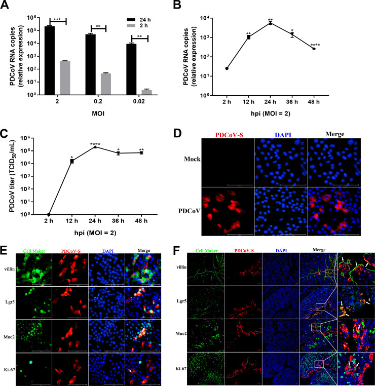
FIG 1.
Porcine intestinal enteroids are susceptible to PDCoV infection. (A) Detection of PDCoV replication in porcine intestinal enteroids by RT-qPCR. Porcine ileal enteroid monolayers were mock inoculated or inoculated with PDCoV at the indicated MOIs for 2 h at 37°C. Enteroid monolayers were harvested at 2 hpi or 24 hpi after removal of virus inoculum and washing of cells three times to remove unattached viruses. Total cellular RNA was extracted, and PDCoV genomes were quantified by RT-qPCR. (B and C) Kinetic curve of PDCoV infection in porcine ileal enteroids. Porcine ileal enteroid monolayers were inoculated with PDCoV at an MOI of 2. The kinetics of PDCoV yielded at the indicated time points postinfection were determined by RT-qPCR (B) or titration (C). The scale data of panels A to C were taken as log(fold change) values and expressed as a power of 10. Each data bar represents the mean of results from three wells for each treatment and time point. Error bars denote deviations. *, P < 0.05; **, P < 0.01; ***, P < 0.005; ****, P < 0.001. (D) Detection of PDCoV infection in ileal enteroids by IFA. The enteroid monolayers were infected with PDCoV at an MOI of 2 for 24 h and then were fixed with 4% paraformaldehyde, and PDCoV S protein expression was confirmed using mouse anti-PDCoV S monoclonal antibody (red). (E and F) Double immunofluorescence labeling of ileal enteroids infected with PDCoV for 24 h (E) or ileal tissues collected from SPF piglets orally inoculated with PDCoV for 72 h (F). Enterocytes, intestinal stem cells, goblet cells, and proliferating cells were stained with anti-villin, anti-Lgr5, anti-mucin 2, and anti-Ki-67 antibodies (green), respectively. PDCoV was labeled with mouse anti-PDCoV S protein monoclonal antibody (red). DAPI-stained nuclei are shown in blue. Arrowheads denote specific cell maker-positive and PDCoV+cells (F). Scale bar, 100 μm in panel E and 50 μm in panel F.

