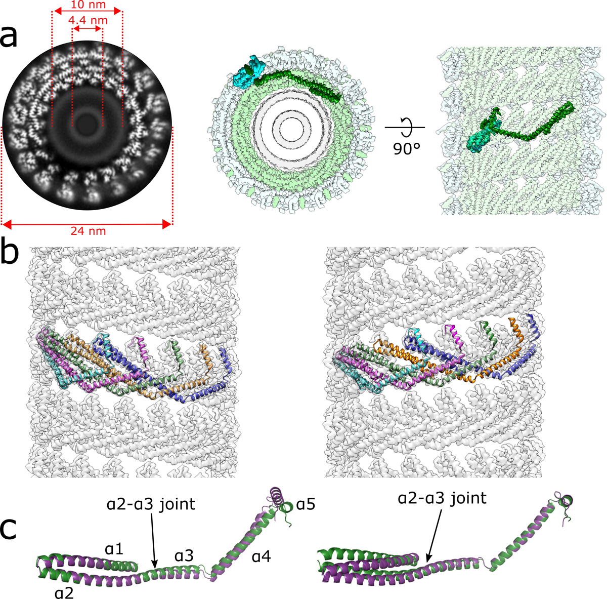
Extended Data Fig. 2. CryoEM reconstruction of the membrane-bound CHMP1B+IST1 filament at higher curvature and comparison of left- and right-handed CHMP1B+IST1 filaments.

a, CryoEM 3D reconstruction of the membrane-bound left-handed CHMP1B+IST1 filament. End-on view down the helical axis in grey-scale (left) or colored (middle). Right, internal view looking outward from the membrane surface along the helical axis. IST1 protomers (cyan) bind to the exterior of CHMP1B (green), leading to constriction of the membrane (grey). IST1 and CHMP1B promoters are highlighted in dark cyan and green, respectively. Diameters of the entire tube and membrane leaflet peak-to-peak distances are annotated. b, Electron density maps of CHMP1B from the left-handed (left) or right-handed (right) membrane-bound CHMP1B+IST1 filaments. Five copies of CHMP1B are shown as ribbons. c, Superposition of a CHMP1B protomer from the left-handed (purple) and right-handed (green) CHMP1B+IST1 filaments aligned to the CHMP1B N-terminal α1-α2 helices (left) or C-terminal α4-α5 helices (right).
