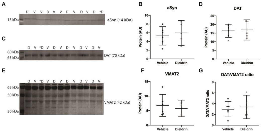
Figure 8: Effect of dieldrin exposure on levels of total α-syn, DAT and VMAT2 in the striatum of male animals.

Monomeric α-syn (A), DAT (C) and VMAT2 (E) were detected by western blot (vehicle: n = 6; dieldrin: n = 5). Samples are in mixed order for more accurate quantification. D = dieldrin, V=vehicle. Dieldrin sample with a * was excluded from all analysis. This sample was not stored properly and ran a typically on some blots. Full blots and total protein staining are shown in Supplementary Figure 6. B) Quantification shows no effect of dieldrin on α-syn levels in the striatum (unpaired t-test with Welch’s correction: p = 0.6279). D) Quantification shows no effect of dieldrin on DAT levels in the striatum (unpaired t-test with Welch’s correction: p = 0.8469). F) Quantification of the 42 kDa band of dieldrin shows no effect of dieldrin on VMAT2 levels in the striatum (unpaired t-testwith Welch’s correction: p = 0.5764). G) Dieldrin shows no effect on DAT:VMAT2 ratio (unpaired t-test with Welch’s correction: p = 0.6700). Data shown as mean+/− 95% CI.
