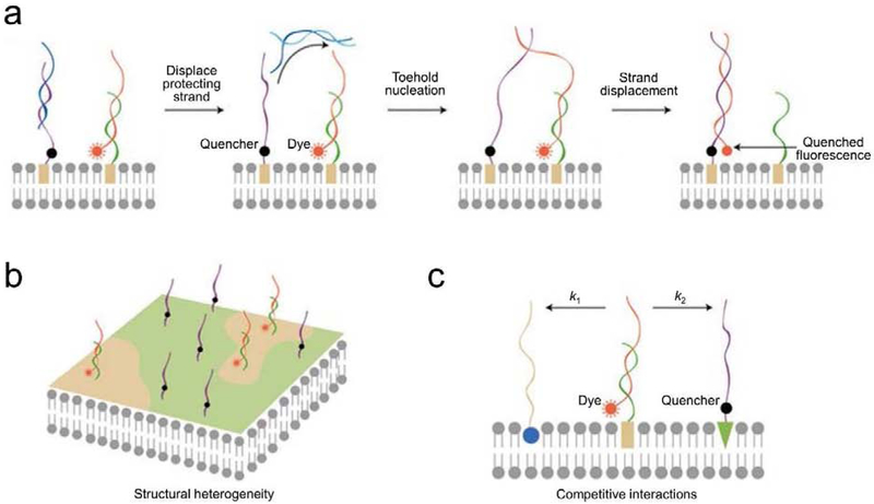
Figure 4.
(a) Schematic of using DNA probes for monitoring transient lipid–lipid encounters on live cell membranes [24]. The addition of an initiator strand (cyan) hybridized and removed the block strand (blue). As a result, once there was a membrane lipid–lipid encounter, the translocation of the DNA probe (red) from one anchor site (green) to another (purple) induced the quenching of fluorescence. (b) Lipid-DNA probes can be used to study membrane heterogeneity. Different lipids preferred to insert into different membrane domains. Lipids of different domains tended to encounter less with each other and resulted in a slow fluorescence quenching. (c) Lipid-DNA probes can be used to measure the competitive interactions among different membrane lipids. Dye-labeled lipid (orange rectangle) was given two possible destinations, unlabeled lipid 1 (blue ball) or quencher-labelled lipid 2 (green triangle). The relative interaction rate (k1 and k2) could be determined based on the rate of fluorescence quenching. Figures are adapted and modified with permission from reference [44].

