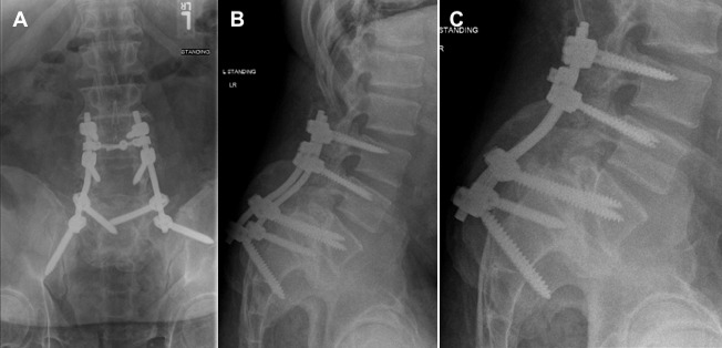
Figure 3.

Three views of circumferential interbody fusion using transsacral fixation in a patient with high-grade spondylolisthesis (HGS). (A) Anteroposterior (AP) radiograph of sacral pedicle – vertebral body (transsacral) fixation. (B) Lateral radiograph of the sacral pedicle – vertebral body fixation. (C) Dedicated lumbar radiograph of the transsacral lumbar fusion from L3-S1.
