
Figure 4.
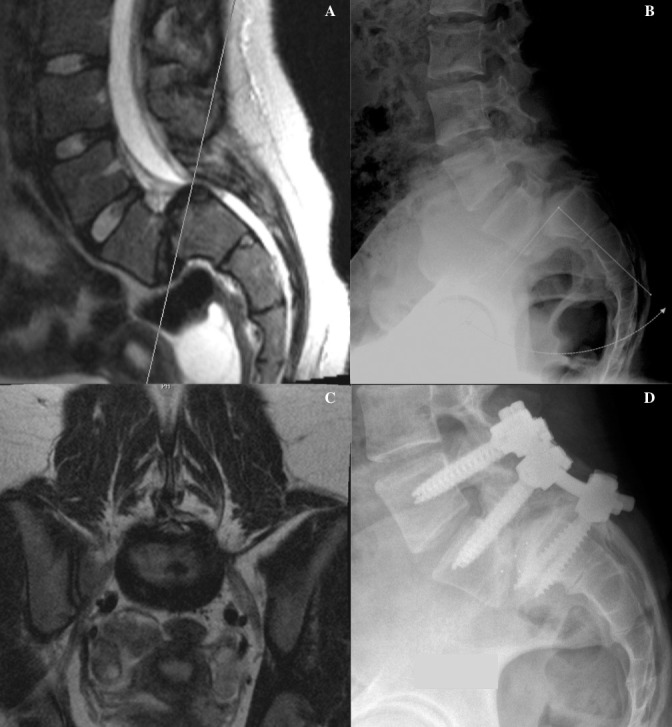
This is a 14-year-old gymnast presented with chronic lower back pain without radicular symptoms. The (A) T2-weighted lumbar magnetic resonance (MR) image and (B) lateral neutral lumbar plain radiograph show exaggerated lordosis and a grade III spondylolisthesis in addition to mild posterior wedging of L5 and mild deformity of the superior S1 end plate. (C) The axial T2-weighted MR image at L5-S1 depicts severe deformity at this level. (D) The patient underwent a laminectomy at L4-S1 with a posterolateral fusion at L4-5 and transforaminal lumbar interbody fusion (TLIF) at L5-S1 following a reduction attempt at L5-S1.