
Figure 3.
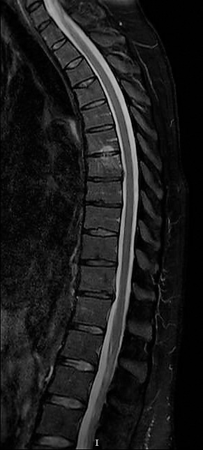
Magnetic resonance imaging (MRI) sagittal T2 at 3-month follow-up—Partial resolution of the Schmorl node (SN) hyperintensity at T7; some novel inferior end plate edema at T6.
Official websites use .gov
A
.gov website belongs to an official
government organization in the United States.
Secure .gov websites use HTTPS
A lock (
) or https:// means you've safely
connected to the .gov website. Share sensitive
information only on official, secure websites.
Magnetic resonance imaging (MRI) sagittal T2 at 3-month follow-up—Partial resolution of the Schmorl node (SN) hyperintensity at T7; some novel inferior end plate edema at T6.