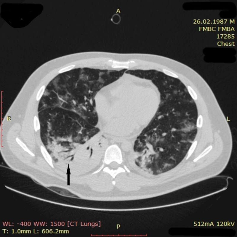
Figure 10. Chest CT imaging of patient 3 on day nine.
Chest CT performed on day nine showed a distinct positive change in CT picture compared to that of day four. The ground-glass opacities noticeably decreased in amount and density all over the lungs. Pneumatized parenchyma could be seen again in all pulmonary segments. The total volume of affected lung tissue was down to approximately 50%. However, a new zone of pulmonary tissue consolidation with a symptom of air bronchogram (black arrow) was found in the lower lobe of the right lung, suggestive of pneumonia
CT: computed tomography

