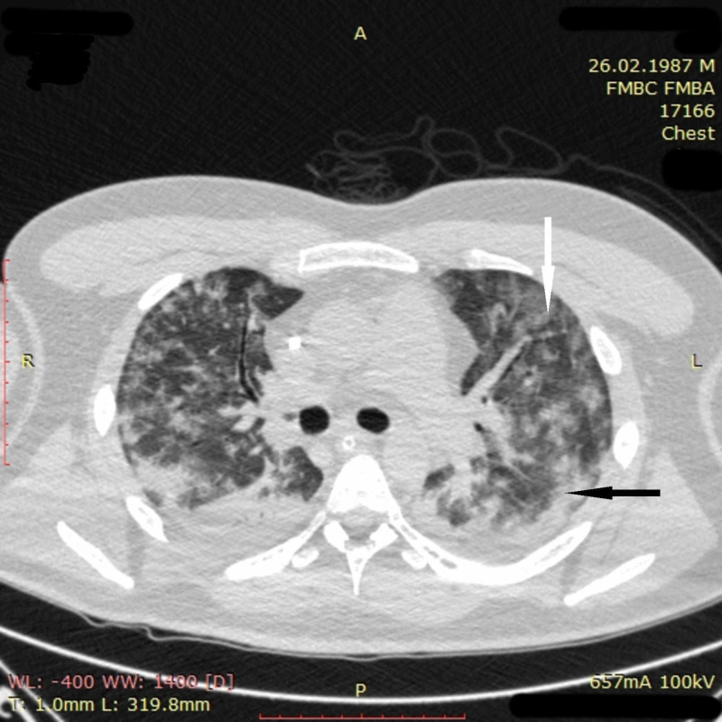
Figure 9. Chest CT imaging of patient 3 on day four.
CT scan on day four demonstrated the appearance of multiple merging ground-glass opacities (white arrow) and areas of consolidation (black arrow) in all segments of the lungs. Partial atelectasis of both lower lobes was also noted. In general, pathological changes involved more than 75% of pulmonary tissue. CT picture was suggestive of ARDS
CT: computed tomography; ARDS: acute respiratory distress syndrome

