Figure 3.
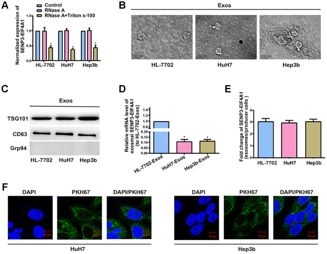
Exosomal SENP3-EIF4A1 mediates intercellular communication. Exos are separated from the medium of HL-7702, Hep3b and HuH7 cells. (A) Detection of the normalized expression of SENP3-EIF4A1 in the medium of HL-7702, Hep3b and HuH7 cells receiving treatment with RNase (2μg/ml) alone or combined treatment with RNase (2μg/ml) and Triton X-100 (0.1%) for 20min via qRT-PCR. (B) Micrographs of exos separated from HL-7702 (left), HuH7 (middle) and Hep3b cells (right, bars=200 nm). (C) Examination of TSG101, CD63 and Grp94 in exos of cell lines via Western blotting. (D) Detection of exosomal SENP3-EIF4A1 of HL-7702, Hep3b and HuH7 via qRT-PCR. (E) Assessment of the fold change of SENP3-EIF4A1 between exos of HL-7702, Hep3b and HuH7 and their producer cells via qRT-PCR. (F) Exos from HL-7702 cells are labeled with PKH67; green represents PKH67, and blue represents nuclear DNAs stained by DAPI. Hep3b and HuH7 cells undergo 3 h of incubation with exos from HL-7702 cells. Results are shown as mean ± SD. *P<0.05. All of the experiments were performed in triplicate.

