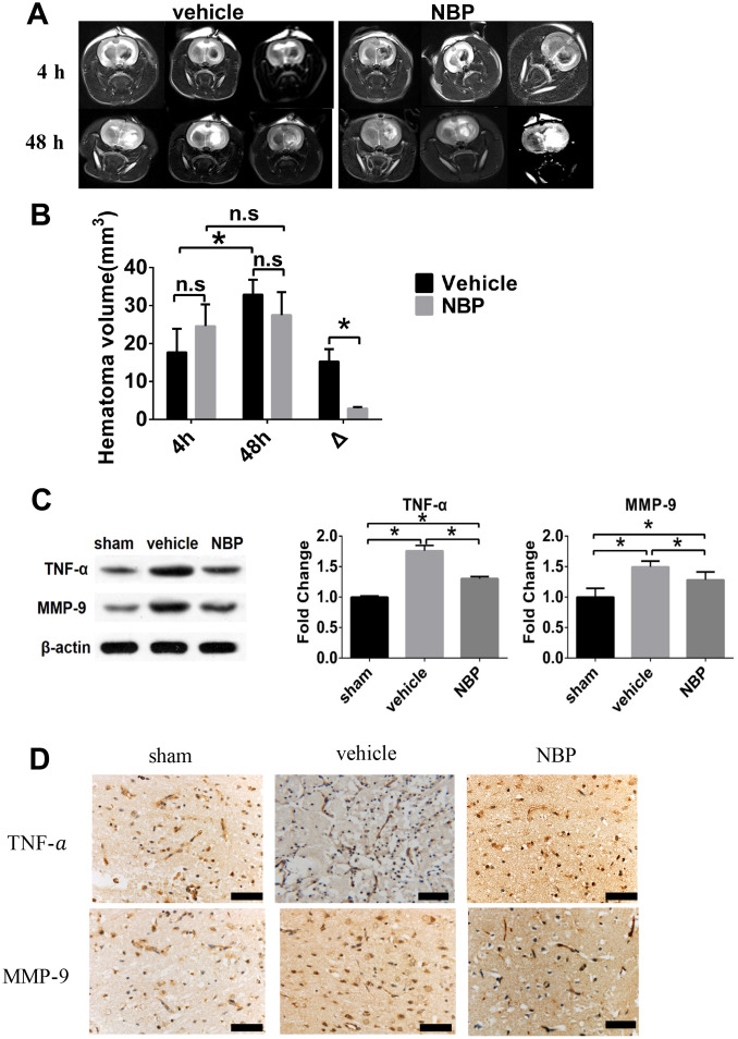
Figure 2.
Effect of NBP on the changes of injured area volume post-ICH. (A) Representative T2-WI images of the vehicle and NBP groups; (B) The hematoma volume at different time points (4h and 48h post-ICH) were quantified; (C) TNF-α and MMP-9 expression in peri-hematoma brain tissue; (D) TNF-α and MMP-9 expression in peri-hematoma brain tissue measured by immunohistochemistry staining (scale: 200 μm). Δ represented the expanded injured area volume, which was calculated by subtracting the volume measured at 48 hours from that at 4 hours. Data are presented as the mean ± SD (n=3, each group). n.s., no significant difference; *, P < 0.05. ICH: intracerebral hemorrhage; NBP: butylphthalide; MMP-9: matrix metalloproteinase-9; TNF-α: tumor necrosis factor-alpha.

