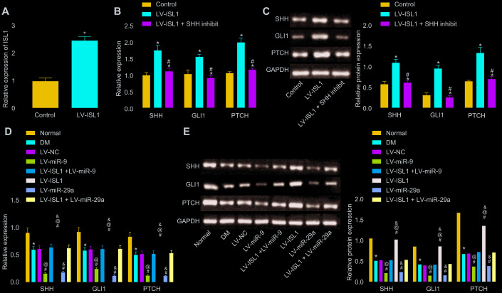
Figure 2.
miR-9 and miR-29a regulate ISL1-mediated SHH signaling pathway. (A) The relative expression of ISL1 after lentiviral infection determined by RT-qPCR. (B) mRNA expression of SHH, GLI1, and PTCH in serum of rats after lentiviral infection detected by RT-qPCR. (C) The protein bands and expression of SHH, GLI1, and PTCH in serum of rats after lentiviral infection normalized to GAPDH detected by Western blot analysis; * p < 0.05 vs. the LV-NC group; # p < 0.05 vs. the LV-ISL1 group. (D) The effect of miR-9 and miR-29a on mRNA expression of SHH, GLI1, and PTCH in serum of rats detected by RT-qPCR. (E) The effect of miR-9 and miR-29a on protein bands and expression of SHH, GLI1, and PTCH in serum of rats normalized to GAPDH detected by Western blot analysis; * p < 0.05 vs. the normal group; # p < 0.05 vs. the DM group; @ p < 0.05 vs. the LV-ISL1 + LV-miR-9 group; & p < 0.05 vs. the LV-ISL1 + LV-miR-29a group. Data were measurement data and expressed as mean ± standard deviation; t test was performed for pairwise comparison; one-way analysis of variance was performed for multiple-group comparison; n = 8; the experiment was repeated three times independently.

