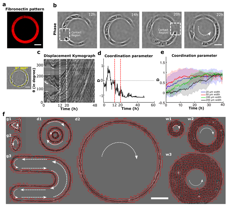
Figure 1. Experimental setup and directed collective migration of MDCK trains.
a, Micro-contact printed pattern of Cy3 conjugated fibronectin in the shape of annular ring (ring outer diameter = 200 μm, track width = 20 μm). b, Phase contrast images of train of MDCK cells after initial seeding and spreading. Dashed white box indicates the contact regions where small cell trains merged into larger trains at 12h and 20h. c, Spatio-temporal displacement of cells traced along the circumferential midline of the ring track. Small train merging event is indicated by red dashed line. d, Evolution of coordination parameter, D, of cell trains in the ring with time (at t=20h cell train in ring begins to rotate in anti-clockwise direction). e, Coordination parameter, D, of cells in ring with different widths: 20 μm (n=62, m=3),50 μm (n=27, m=2),100 μm (n=8), 200 μm (n=8) respectively, shows a collective migration of cell trains independent of track width. f, Velocity vectors representing directed collective cell migrations for a variety of confined geometries (g1-triangle, g2-ellipse, g3-U shape), diameter (d1:outer-400 μm, middle-200 μm, inner-100 μm, d2-1mm), and width (w1-50 μm, w2-100 μm, w3-200 μm). All error bars indicated are standard deviations. Scale bars a, b, c: 50 μm and f: 200 μm.

