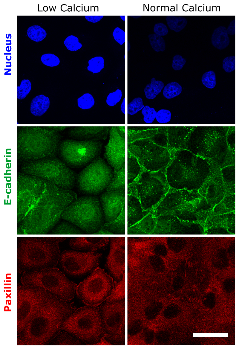
Extended Data Figure 8. Cell-cell junctions and focal adhesions in the presence of low and normal calcium media.
Nucleus (blue)-E-cadherin (green)-paxillin (red) immunofluorescence of cell monolayers. E-cadherin accumulation at cell-cell contacts detected in normal medium, disappear in low calcium. By contrast, paxillin staining reveals the presence of focal adhesions in both conditions. Scale bar: 20 μm.

