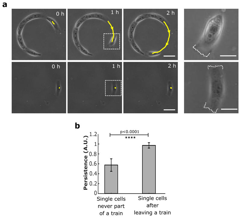
Extended Data Figure 9. Single cell migration persistency in the absence of cell-cell junctions.
a, Top panel: Phase contrast images of cell trains at low density that show spontaneous detachment of single cells at the free edges. Sequential images showing cell detachment followed by a polarized persistent migration. Expanded view of the dashed white box (last column) reveals the active lamellipodial activity on one side of the cell (marked with white dashed line). Bottom panel: Control experiment showing the behavior of a single MDCK cell with no previous contact with other cells. No preferential migration is observed. Both edges of the cell show lamellipodial activity as marked by dashed white line in the expanded view (last column) from white dashed square box. (Scale bars: 20 μm). The yellow lines indicate the distance travelled by the cells at given times. b, Persistence of cell movements for single isolated cells (n=12) and for single cells detaching from sub-confluent trains (n= 11). All error bars indicated are standard deviations. All scale bars unless mentioned specifically: 50 μm.

