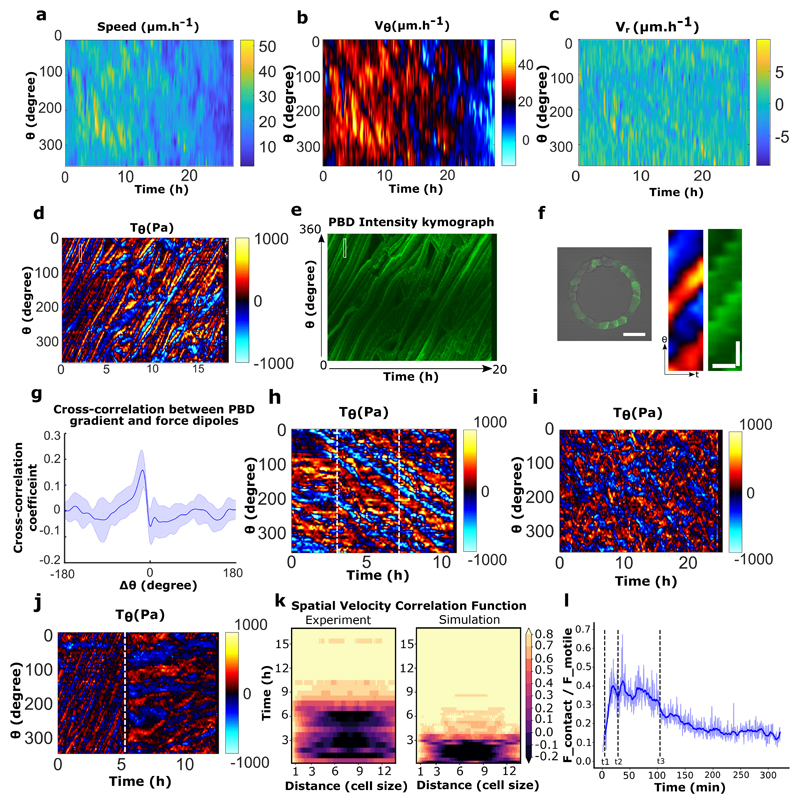
Extended Data Figure 10. Spatio-temporal distribution of velocities and forces under various condition.
a, Spatio-temporal kymograph of cell train speed for the experiment referred in Fig.3d. b-c, Spatio-temporal tangential speed and radial speed kymograph for the experiment referred in Fig.3d. d, Tangential traction force spatio-temporal profile of MDCK-PBD cells in rotating ring. e, Corresponding PBD intensity profile and tangential traction force distribution as shown in panel d. f, MDCK-PBD cells on ring pattern (Left). Expanded view of corresponding regions of interest for PBD gradients and traction force dipoles (marked by white rectangular box in panel d, e) (right). Scale bar, Ө: 10 μm, t: 30 minutes. g, Cross correlation of tangential traction force profile and PBD signal show a high spatial correlation (n=10,m=2). h, Tangential traction forces post EGTA treatment indicate single cell level force dipoles in a rotating ring before and after EGTA treatment (beginning of rotation is marked by the white dashed line ~ at 3h), EGTA addition is marked by the white dashed line at ~7h. i, Tangential traction force distribution in α-catenin KD cells. j, Tangential traction force pattern before and after Apr2/3 inhibition. The white dashed line marks CK666 drug addition (100 μM). k, Spatial velocity correlation C(x’,t) as a function of distance x’ in experiments and simulations (both with 15 cells) showed similar migration dynamics. where x, x’ are curvilinear abscissas, u is the 2 angular velocity (positive in counter-clockwise direction) and t is time. l, Ratio of contact and motile forces in 1 simulation (taken from Fig. 4b), recapitulating the force-distribution transition observed in Fig. 2f-h: before polarization starts (t<t1), and once collective rotation is established (t>t3), cells migrate as single dipole and their viscous interaction with the substrate is mainly balanced by their motility. During the coordination process (between t1 and t3), cells form larger dipoles and the contact forces contribute to viscous force balance (t1: cells start polarizing, until they are all polarized at t2, and rotate collectively from t3) (blue thick line: smoothing over time). All error bars indicated are standard deviations. All scale bars unless mentioned specifically: 50 μm.

