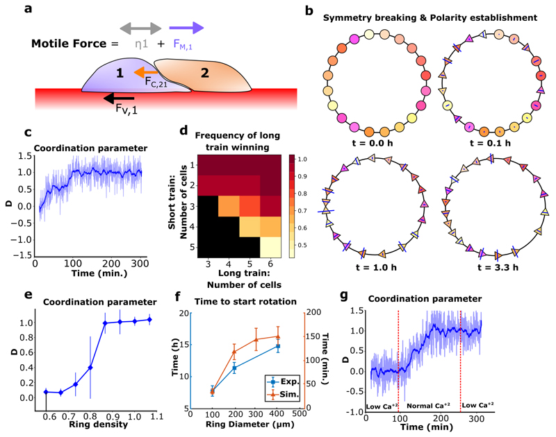
Figure 4. Mechanics-based simulations of epithelial cell migration on a ring.
a, Schematic of force balance in epithelial cell train. The forces are FV (viscous), FC (contact), FM (directed motile force) and the diffusion ŋ. b, Coordination emergence in a simulation. Circles: centers of non-polarized cells. Triangles: centers of polarized cells, pointing in their polarity direction (cell boundaries are not represented). Blue lines indicate the intensity of contact forces on a cell. c, Simulations reveal that persistent coordination emerges over time (blue pale line: coordination parameter for n=1 simulation, smoothed over time in the blue thick line). d, Colormap of the probability for the longer train to "win" a collision versus the number of cells in both trains. Black square: no data. n=20. e, Coordination parameter in simulations after 1000 timesteps (~400 minutes, averaged over last 8 minutes) versus cell density demonstrates that confluent density is necessary to achieve coordination. n=20 per point. f, Coordination time in experiments (n=20) and simulations (n=25) with varying ring sizes. g, Simulations varying the contact stiffness parameter over time to modulate cell-cell interactions (blue pale line: coordination parameter for n=1 simulation, smoothed over time in the blue thick line). All error bars indicated are standard deviations. * ring diameter is 200 μm unless stated otherwise.

