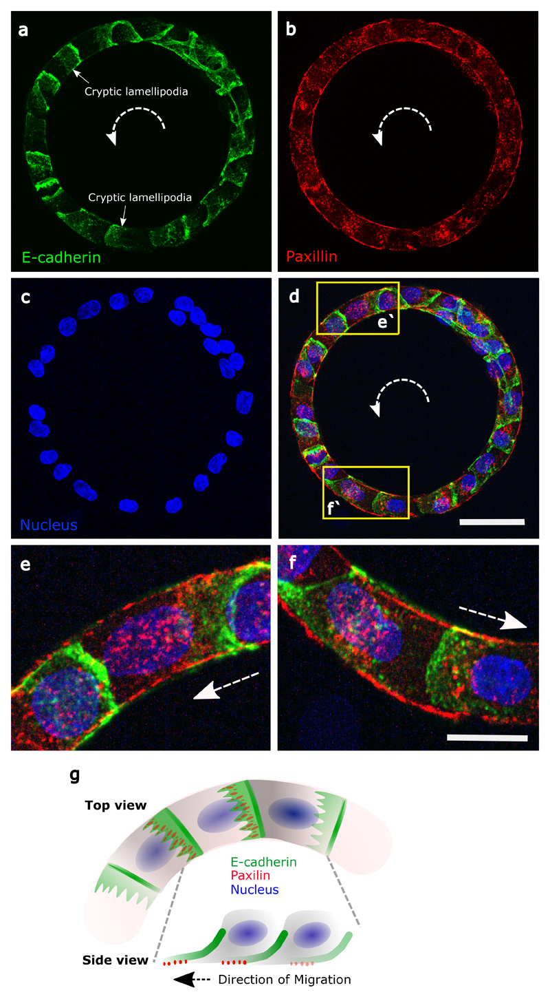
Extended Data Figure 3. Immunostainings of cell-substrate adhesions (paxillin) and cell-cell adhesion (E-cadherin).
a, Immunofluorescence for E-cadherin-GFP (Z-projection) in a rotating ring. Anti-clockwise direction is indicated by the white dashed arrows. The diffused E-cadherin distribution indicates the cryptic lamellipodia. b, Basal immunofluorescent paxillin staining labelled focal adhesions. c, Nuclei labelled in blue. d, Merge E-cadherin (green), paxillin (green) and nuclei (blue). (e-f) Images showing enlarged views of (e'-f'). g, Schematic showing single cells in rotating ring with cryptic lamellipodia revealed by polarized distributions of E-cadherin and paxillin. Scale bars: 50 μm (a-d), 20 μm (e-f)

