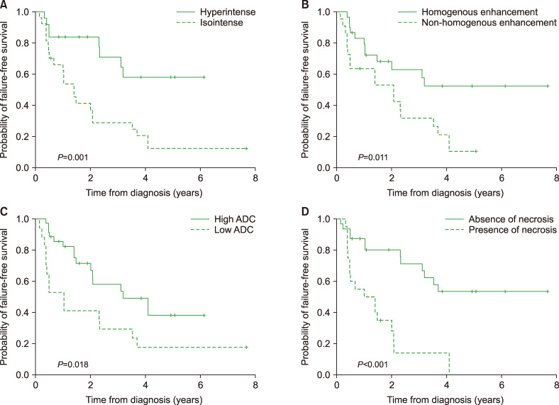
Fig. 2.
Kaplan-Meier curves for failure-free survival (FFS). Patients with hyperintense signal on T2-weighted imaging (P=0.001) (A) and homogenous enhancement (P=0.011) (B) had better FFS, while patients with low ADC (P=0.018) (C) and necrosis had poor FFS (P<0.001) (D).
Abbreviation: ADC, apparent diffusion coefficient.

