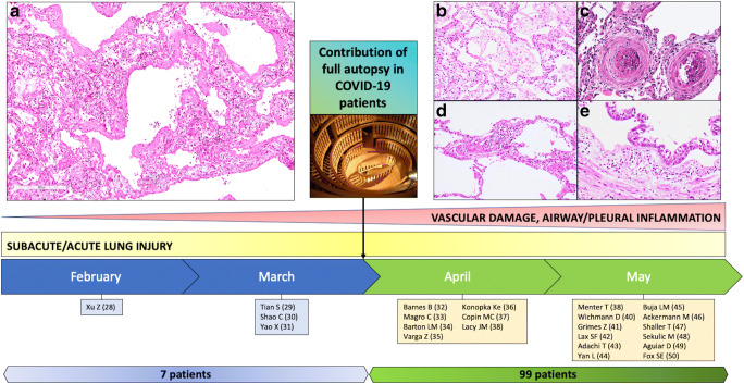
Fig. 2.
Timeline of autopsy studies focusing on lung lesions in COVID-19 patients. Since the end of March, the increased number of full autopsies has led to a better knowledge of the pathophysiology of the disease. Together with the features of acute lung injury, vascular involvement has been reported. a, b Acute lung injury: hyaline membrane in alveolar space (hematoxylin and eosin stain, original magnification a × 100; b × 200). c, d Vascular damage: two microthrombi in lung small vessels (hematoxylin and eosin stain, original magnification × 200), capillary inflammation (hematoxylin and eosin stain, original magnification × 200). e Airway inflammation: tracheal section showing a polymorphous inflammatory infiltrate of the submucosal layers (hematoxylin and eosin stain, original magnification × 200)

