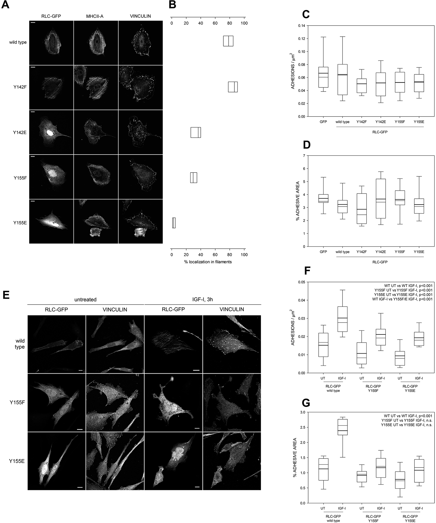
Figure 2. Y155 is required for the correct localization of RLC to actomyosin filaments and adhesion elongation.

(A) Images of fibronectin-bound, CHO.K1 cells transfected with the indicated GFP-tagged versions of RLC. Bars=10 μm. Pictures are representative of >500 cells examined in three independent experiments.
(B) Quantification of the filamentous localization of the RLC mutants shown in (A). Boxes represent the 25th and 75th percentile of three independent experiments in which >500 cells were analyzed. p<0.01 between wild type/Y142F and the other conditions; also between Y155E and the other conditions.
(C-D) Quantification of the number of adhesions per μm2 (C) and total percentage of the area of the cell that corresponds to focal adhesions, that is, adhesive area (D). Boxes represent the 25th and 75th percentile and whiskers correspond to the 10th and 90th percentile of three independent experiments in which n ≥ 20 cells (>2000 adhesions) were analyzed. Throughout the study, thick line represents the mean and thin line the median. There are no statistically significant differences among conditions.
(E) Images of fibronectin-bound, CHO.K1 cells transfected with the indicated GFP-tagged versions of RLC. After 2h of adhesion, cells were starved overnight and then stimulated with IGF for 3h. Bars=10 μm. Pictures are representative of >200 cells from three independent experiments.
(F-G) Quantification of the number of adhesions per μm2 (F) and adhesive area (G) in the conditions represented in (E). n ≥ 20 cells from three separate experiments (>2000 adhesions per condition). The significances of the relevant comparisons between conditions are shown on the top right corner of each panel. WT stands for wild type throughout the study.
