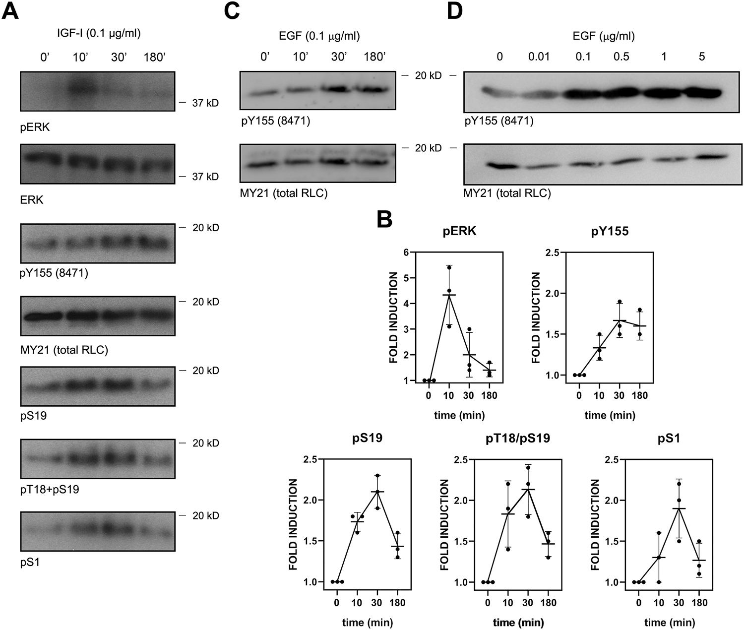
Figure 6. Growth factor-dependent phosphorylation of RLC on Y155 is kinetically different from its phosphorylation on S1, S19 and T18/S19.

(A) CHO.K1 cells were serum-starved for 16h, then treated with IGF-I (0.1 μg/ml) for the indicated times, lysed with Laemmli buffer and blotted for the indicated antigens. Experiment is representative of three performed.
(B) Quantification of the densitometric profiles of the blots shown in (A) and its biological replicates.
(C) Serum-starved A549 cells were treated for the indicated times with 0.1 μg/ml EGF and processed as in (A). Experiment is representative of three performed.
(D) Serum-starved HEK-293 cells were treated for 30 min with the indicated dose of EGF. Experiment is representative of three performed.
