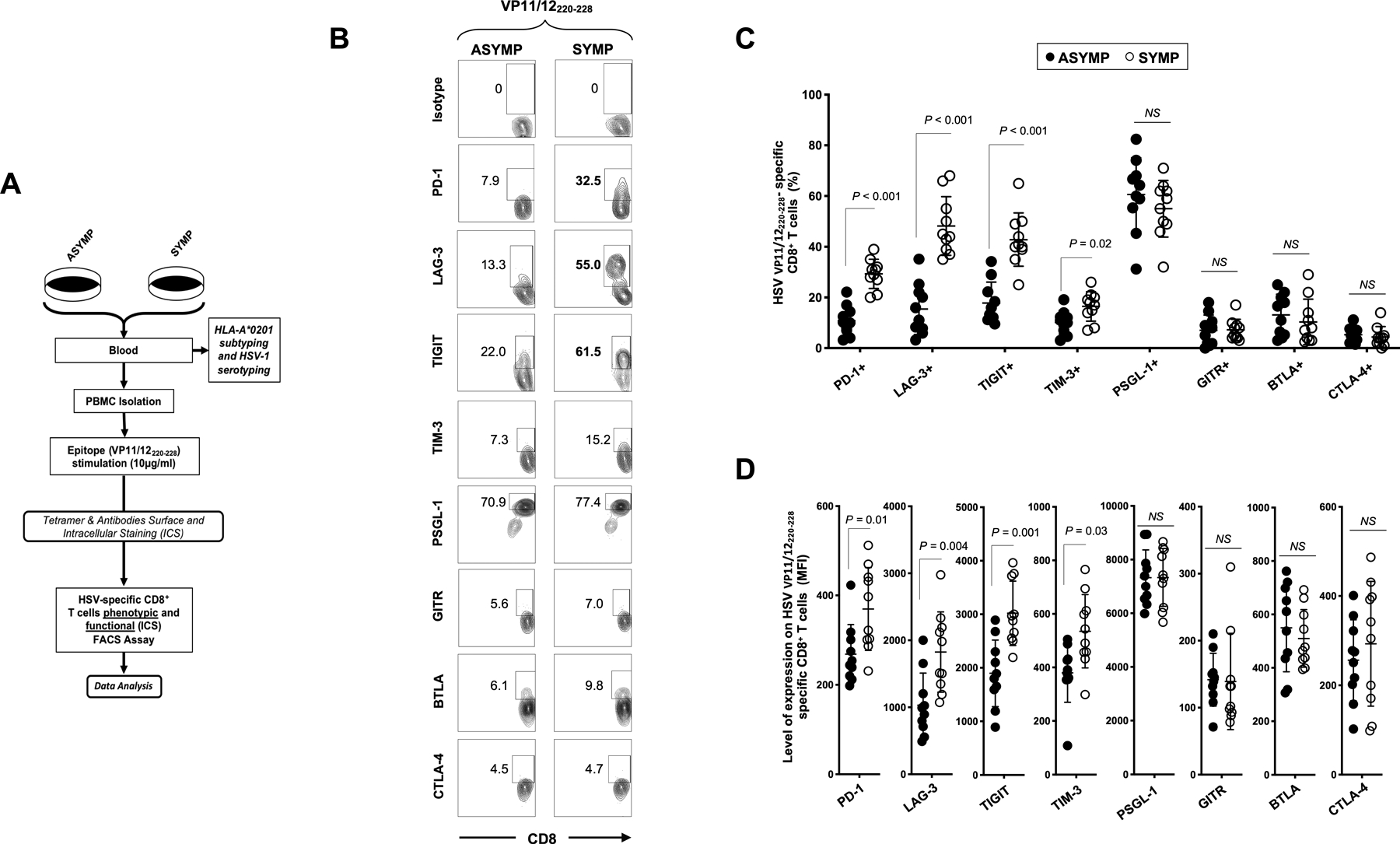
Figure 2: Cell surface expression of exhaustion receptors by HSV VP11/12220–228 epitope-specific CD8+ T cells from symptomatic vs. asymptomatic individuals:

(A) Experimental design for differentially expressed exhaustion molecules detected by FACS on the surface of CD8+ T cells sharing the same HSV-1 epitope-specificities, from SYMP and ASYMP individuals. PBMCs from 10 SYMP and 10 ASYMP individuals were stained for CD3/CD8, VP11/12220–228 tetramer and different exhaustion markers or the isotypic control with the same fluorochrome. The gating strategy is shown in Fig. S1. (B) Representative FACS plots and (C) average frequencies of HSV-1 VP11/12220–228-specific CD8+ T cells expressing PD-1, LAG-3, TIGIT, TIM-3, PSGL-1, GITR, BTLA and CTLA-4 receptors, detected from SYMP vs. ASYMP individuals. (D) Levels of expression of the eight exhaustion receptors on HSV-1 VP11/12220–228-specific CD8+ T cells from SYMP and ASYMP individuals, depicted as Mean fluorescent Intensity (MFI). Each staining was performed in duplicate. The data are expressed as means +/− the standard deviations (SD). The indicated P values, determined using unpaired t test, compared expression of exhaustion receptors between SYMP and ASYMP individuals.
