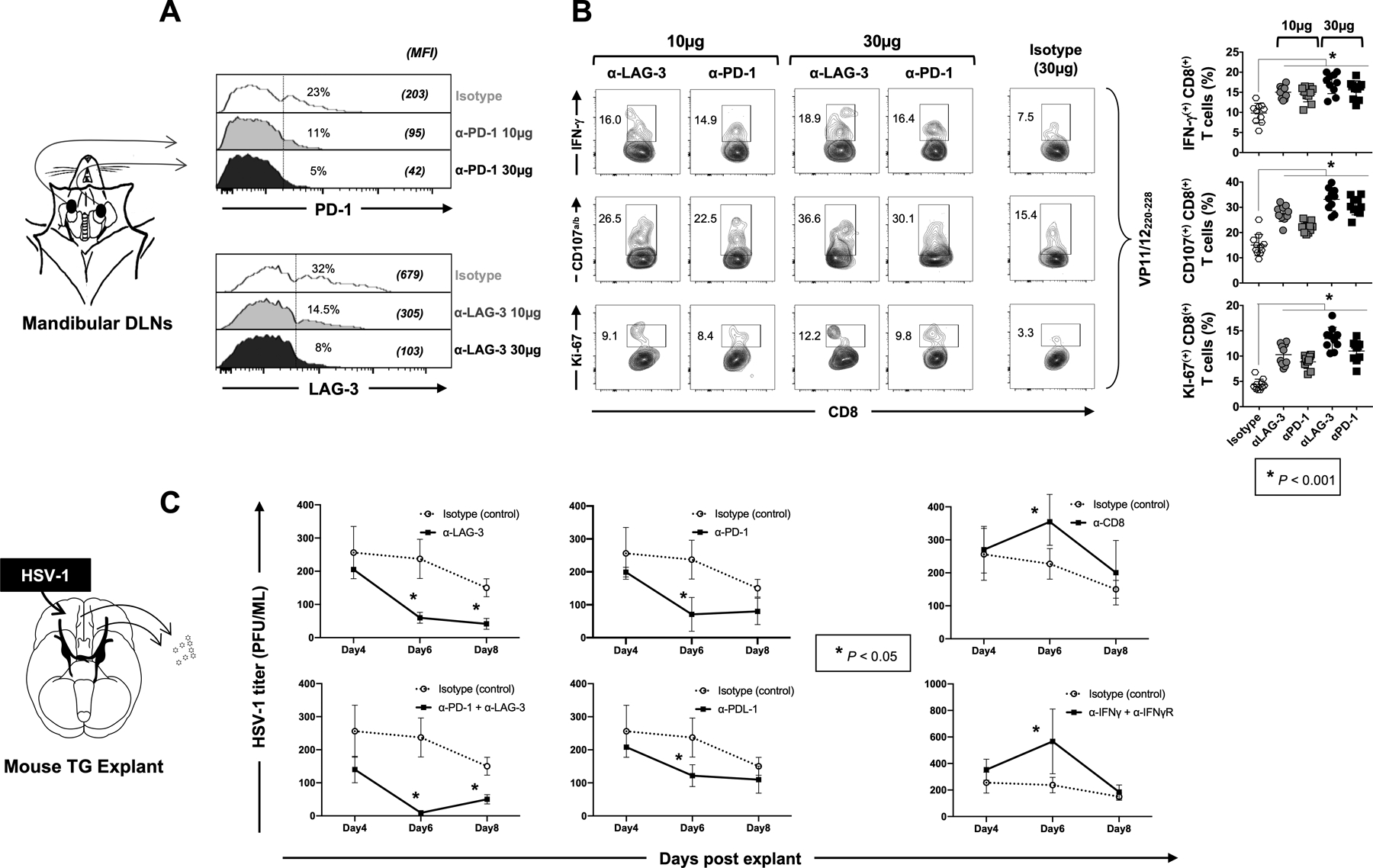
Figure 6: Effect of ex vivo blockade of PD-1, LAG-3 and/or TIM-3 immune checkpoints on function of HSV-specific CD8+ T cells and virus reactivation from TG explants of HSV-1 infected HLA-A*0201 transgenic mice.

HLA-A*0201 Tg mice (n =10 per group of blockade treatment) were ocularly infected with HSV-1 (2.5 × 10⁵ pfu per eye, strain McKrae). Fourteen days post-infection CD8+ T cells derived from submandibular lymph nodes were stimulated for 12 hours with 10 μg/mL of VP11/12220–228 peptide (“RLNELLAYV”). Blocking α-LAG-3 and/or α-PD-1 mAbs were added in the culture for two additional days (at either 10 μg/mL or 30 μg/mL). (A) Representative histograms display the levels of expression of LAG-3 or PD-1 depicted by the MFI on HSV-1 VP11/12220–228-specific CD8+ T cells from HSV-1 infected HLA-A*0201 Tg mice following blockade of LAG-3 or PD-1 immune checkpoints. (B) Representative FACS plots of the percentages of functional HSV-1 VP11/12220–228-specific IFN-γ+CD8+, CD107a/b+CD8+ and Ki-67+CD8+ T cells following in vitro blockade of LAG-3 or PD-1 immune checkpoints. (C) Fourteen days post-infection, cell suspensions from TG explants were incubated in duplicates with 100 μg/mL of α-LAG-3 and/or α-PD-1 blocking mAbs or isotype control mAb. Graphs show the level of reactivated virus (expressed in PFU/mL) detected in the supernatant of TG explants on 4, 6- and 8-days post-incubation. The data are expressed as means +/− the standard deviations (SD). The indicated P-values, calculated using the unpaired t test, show the statistical significances between the control (Isotype) and the different blocking conditions. The results showed here are representative of two independent experiments.
