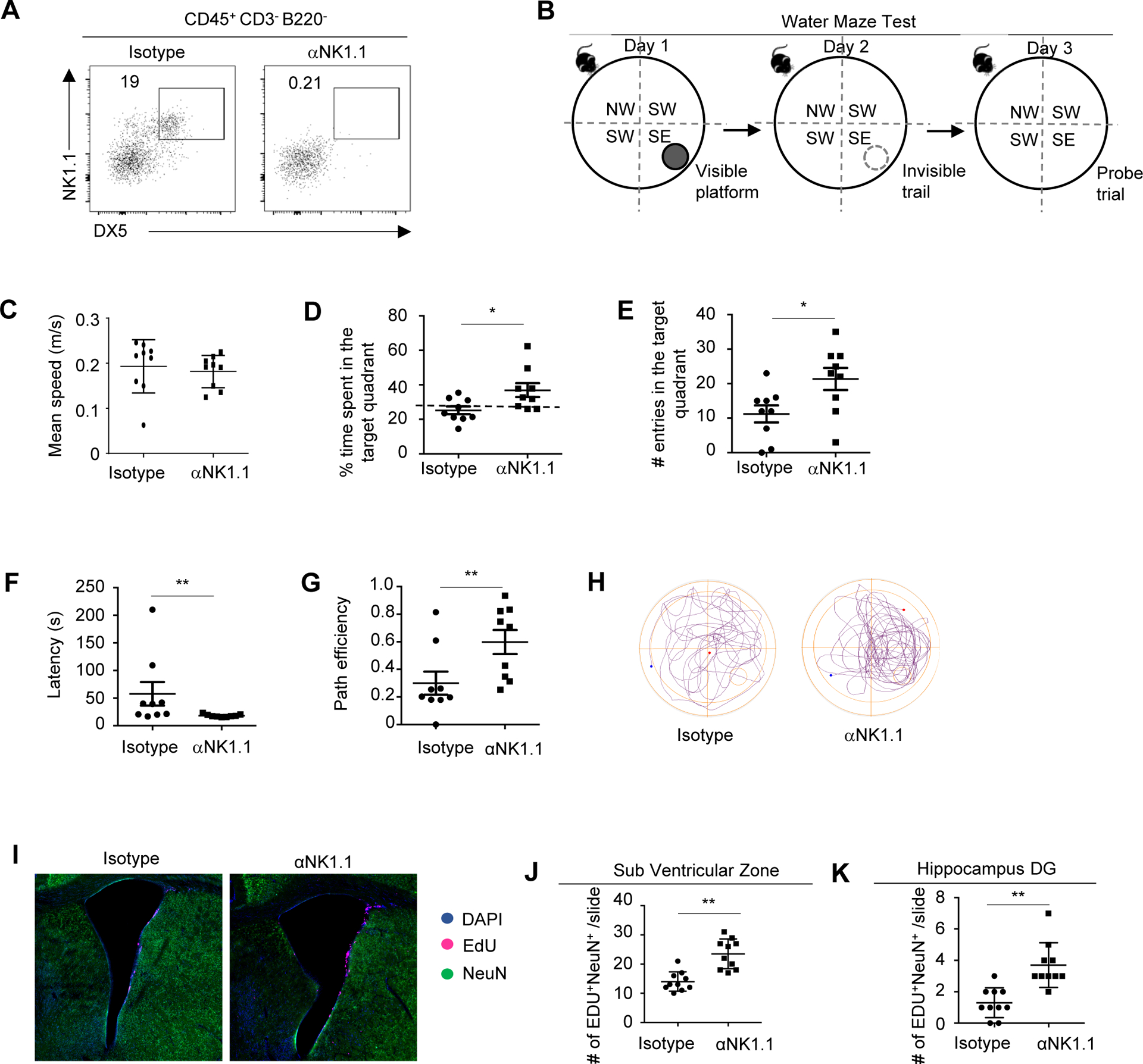
Figure 2. Depletion of NK cells improved cognitive function in 3xTg-AD mice.

A, Representative flow cytometry profiles of NK cells in 7–8 month old 3xTg-AD mice treated with anti-NK1.1 antibodies or isotype controls. Plots were pre-gated on brain CD45+CD3−B220− lymphocytes. B, Experimental scheme of Water Maze Test. C, Mean swimming speed of 7–8 month old 3xTg-AD mice treated with anti-NK1.1 antibodies or isotype controls in the probe trial. D, Time spent in the target quadrant by 3xTg-AD mice treated with anti-NK1.1 antibodies or isotype controls in the probe trial. E, Numbers of entries into the target quadrant in the probe trial. F, Latency to enter the target quadrant in the probe trial. G, Path efficiency of entering the target quadrant in the probe trial. H, Representative path profiles in the probe trial. I. Representative immunofluorescence imaging depicting Edu+ neurons in the SVZ region. J, Numbers of EdU+ neurons in SVZ regions. K. Numbers of EdU+ neurons in hippocampus dentate gyrus regions. DG, dentate gyrus. Data represents 3 independent experiments (A); or are from 9 mice per group and represent two independent experiments (B-H); or from 10 mice per group pooled from two independent experiments (I-K). *p<0.05; **p<0.01.
