
Figure 1.
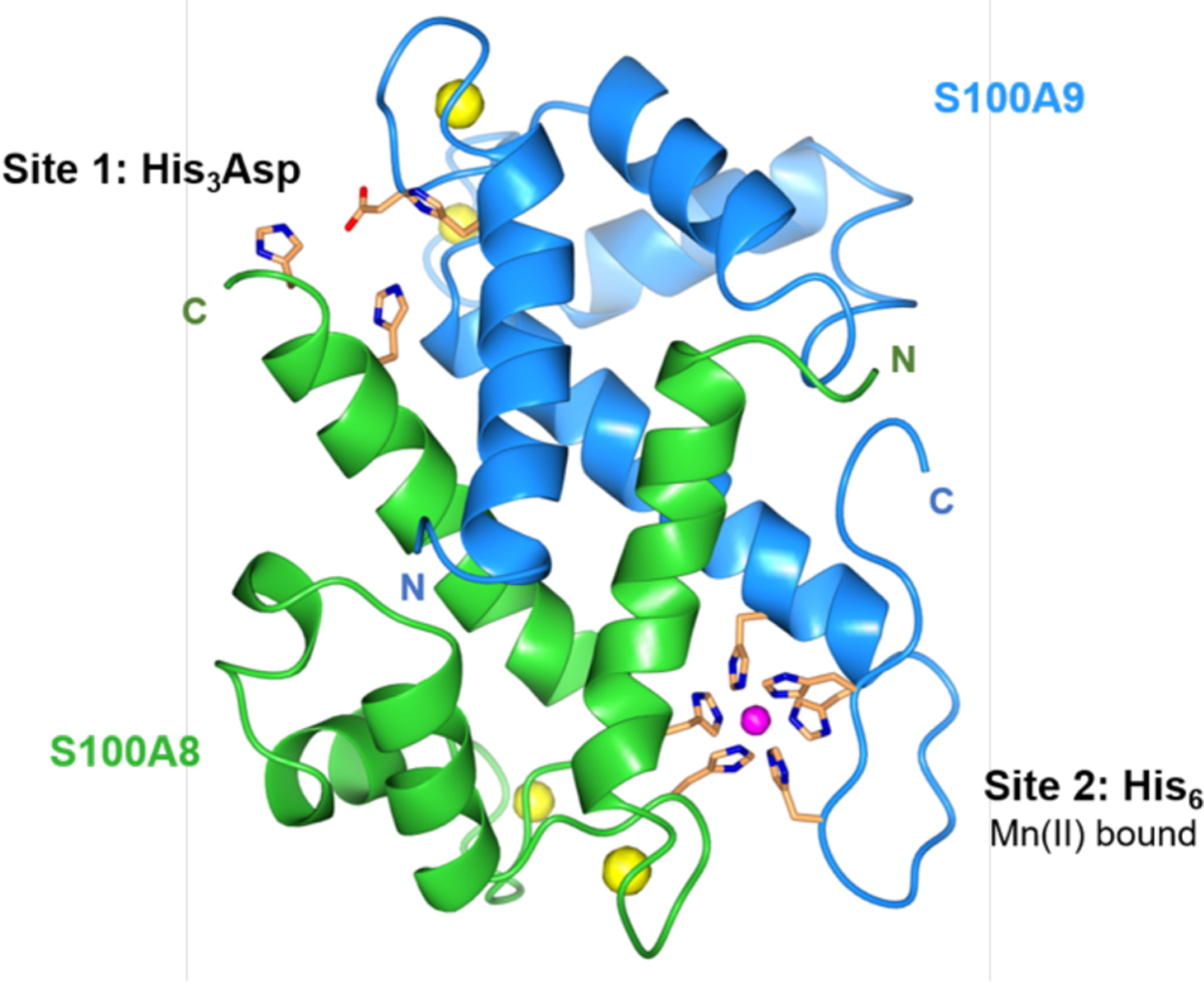
Crystal structure of Mn(II)-, Ca(II)-, and Na(I)-bound CP-Ser (PDB 4XJK).21 A heterodimer unit of S100A8 (green) and S100A9 (blue) is taken from the structure of the heterotetramer. The N- and C-termini of S100A8 and S100A9 are labeled. Mn(II) is shown in magenta; Ca(II) and Na(I) are shown in yellow. The transition-metal binding residues are shown in orange.