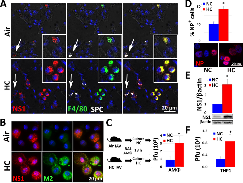
Fig. 2: Hypercapnia increases viral protein expression and viral replication in alveolar macrophages following IAV infection of mice and in IAV-infected human THP-1 macrophages.
Mice were pre-exposed to air or normoxic hypercapnia (10% CO2/21% O2, HC) and infected with IAV (30 pfu), as in Fig. 1A. Animals were sacrificed 4 dpi, and lung tissue sections were stained for viral NS1 (red), F4/80 (green, MØs), SPC (white, AT2 cells) and nuclei (blue); inserts show enlarged view of AMØs, white arrows indicate AT2 cells, n=4–6; results representative of at least 2 independent experiments (A). AMØs obtained by BAL 1 dpi were stained for viral NS1 (red), M2 (green) and nuclei (blue), n=6; results representative of 3 independent experiments (B), or cultured under normocapnic (5% CO2/95% air, NC) or hypercapnic (15% CO2/21% O2/64% N2, HC) conditions for 18 h, after which viral titers in culture supernatants were determined by plaque assay, mean ± SEM, n=6 from 3 independent experiments (C). Differentiated THP-1 MØs were pre-exposed to NC or NC for 2 h, infected with IAV (MOI 2), and cultured in NC or HC for an additional 18 h. THP-1 MØs were then stained for viral NP (red) and the percentage of NP-positive cells was determined (D) or lysed for determination of viral NS1 expression by immunoblot (E) and viral titers in culture supernatants were determined by plaque assay (F); mean ± SEM, n = 4 from 4 independent experiments. *P<0.05 vs. NC + IAV.

