Figure 1.
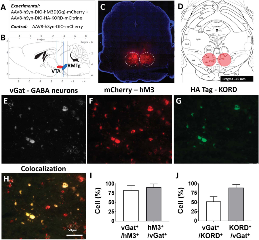
Validation studies demonstrate localization and functionality of RMTg-targeted hM3 DREADDs and KORD. (A) Summary of cre-dependent viral constructs delivered to the RMTg. (B) Orientation and proximity of the RMTg in relation to the VTA. (C) AAV-hsyn-DIO-hM3D(Gq)-mCherry expression in the RMTg of a representative vGat-cre mouse (see also Fig. S1, available online as supplemental digital content at http://links.lww.com/PAIN/A842). (D) RMTg target location on the mouse atlas (−3.9 mm anterior/posterior, ±0.4 mm lateral, and −4.8 mm dorsal/ventral to the bregma). (E) Fluorescence in situ hybridization (FISH) staining of the vesicular GABA transporter (vGat) in RMTg neuronal cell bodies in a representative mouse. (F) hM3D-mCherry expression in the same region as (E). (G) HA tag (KORD) expression in the same region as (E). (H) Overlayed image of (E, F, and G) showing colocalization of hM3D and KORD expression in vGat-expressing neurons of the RMTg; scale bar, 50 μm. (I) 91 ± 6% of red-labelled hM3D+ transcripts in the RMTg colocalized with white, Cy5-labeled neurons expressing vGat RNA, and 83 ± 8% of white Cy5-labelled vGat-cre–expressing neurons colocalized with red, mCherry-labelled hM3D+ transcripts. (J) 90 ± 9% of green-labelled KORD+ transcripts in the RMTg colocalized with white, Cy5-labeled neurons expressing vGat RNA, and 52 ± 13% of white Cy5-labelled vGat-cre–expressing neurons colocalized with green-labelled KORD+ transcripts. RMTg, rostromedial tegmental nucleus; VTA, ventral tegmental area.

