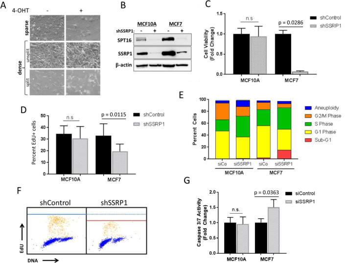
Fig. 1. FACT is essential for the viability of proliferating tumor cells, but not of growth-arrested transformed cells or growing non-tumor cells.
a The effects of Ssrp1 KO on growing and growth-arrested transformed mouse fibroblasts from Ssrp1fl/fl; CreERT2+/+ mice. Microphotographs of cells plated at two different densities, sparse (~10% confluency) in 10% FBS or dense (100% confluency) in 1% FBS, and grown in the presence or absence of 4-OHT for 5 days. Images were taken at the end of 4-OHT treatment for sparse cells and 10 days posttreatment for dense cells, which were either split or not 3 days before imaging. b–g Human mammary epithelial cells, immortalized MCF10A or tumor MCF7, were evaluated 72 h after transduction with lentiviral shRNAs or transfection with siRNAs as indicated, except c. b SSRP1 knockdown (KD) in MCF10A and MCF7 cells. Immunoblotting of cell lysates (“−” shControl). c Effect of SSRP1 KD on MCF10A and MCF7 cells viability. Cells were sparsely seeded 72 h post-transduction, allowed to grow for 10 days, and then stained with methylene blue. Data are presented as the mean ± SD (n = 3) normalized to the corresponding shControl. d Effect of SSRP1 KD on DNA replication of MCF10A and MCF7 cells. EdU incorporation was quantified using immunofluorescence imaging. Data are presented as the mean ± SD (n = 3). e Quantitation of cell cycle distribution using flow cytometry. f Cell cycle analysis using co-staining with EdU and Hoechst (yellow dots, EdU+ cells; blue dots, EdU− cells). Blue and red lines represent the maximum intensity of EdU in shControl and shSSRP1, respectively. g Caspase-3 and -7 activity. Data are presented as the mean ± SD (n = 3).

