
Fig. 19.
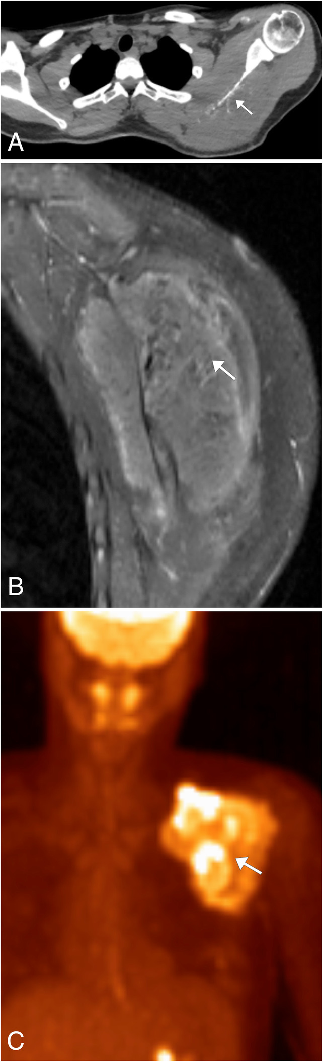
Ewing sarcoma. a Axial noncontrast chest CT demonstrates permeative destructive changes of the left scapula (arrow) with the large soft tissue component of the mass not well seen. b Sagittal T1WI MR image post contrast allows for visualization of the associated enhancing mass (arrow) which involves the surrounding left scapular soft tissues and musculature. c Coronal FDG-PET/CT image demonstrating the hypermetabolic nature of the lesion associated with the left scapula (arrow)