
Fig. 2.
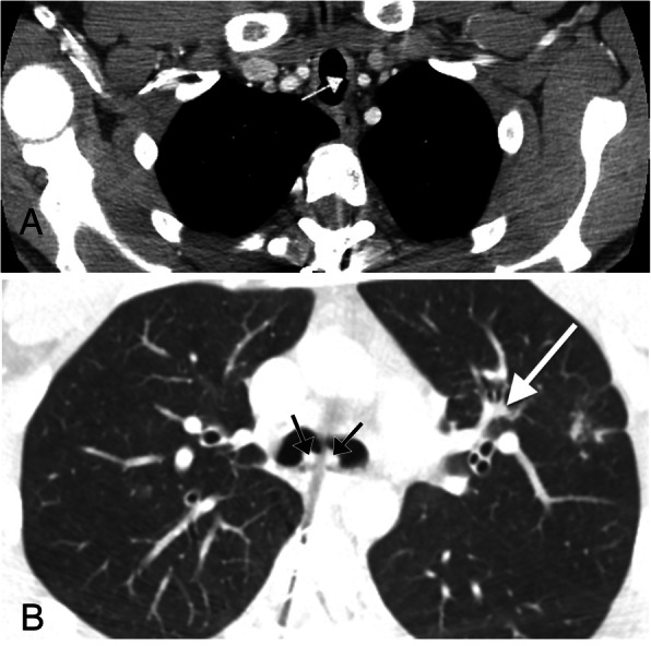
Recurrent respiratory papillomatosis. a Axial chest CT showing a soft tissue attenuation polyp in the lateral wall of the upper thoracic trachea (arrow). b Axial CT image of the same patient at the level of the carina showing two additional soft tissue attenuation polyps (black arrows). This image also demonstrates bronchiectasis in the left upper lobe which occurred secondary to luminal obstruction of a distal airway secondary to a polyp (white arrow)