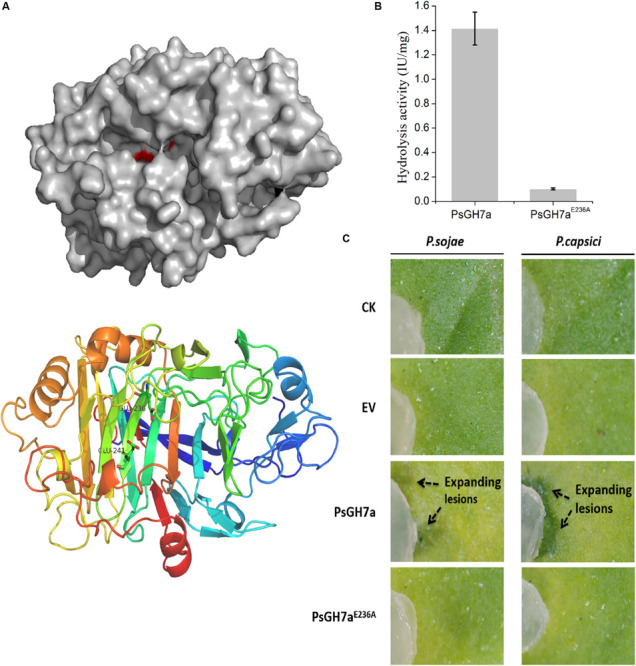
FIGURE 4.

The hydrolysis of PsGH7a promotes Phytophthora invasion. (A) Predicted three dimensional structure of the PsGH7a protein obtained using the homologous Phanerochaete chrysosporium cellobiohydrolase Cel7D as the template (PDB: 1z3t). The color gradient shows the sequence from the N terminus (blue) to the C terminus (red). In particular, the putative catalytic residues Glu236 and Glu241 are presented as green sticks. (B) Cellulase activity (IU) of the wild-type and the mutant form PsGH7aE236A. One unit (U) of cellulase activity was defined as the amount of cellulase that catalyzed the liberation of reducing sugar equivalent to 1.0 μg glucose/min under assay conditions. Three independent biological replicates were used for each protein. (C) After the PsGH7a and the mutant form PsGH7aE236A proteins were infiltrated into N. benthamiana leaves, P. sojae and P. capsici hyphal plugs were put on the infiltration sites. The buffer was infiltrated as a control (CK); the cell-free supernatant from Pichia pastoris GS115 which contains a empty vector (EV) was also infiltrated as another control. Arrow indicates enlarged lesion area. The pictures were taken at 3 days after inoculation.
