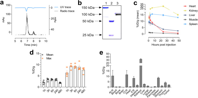
Fig. 1.
a Representative HPLC chromatogram of [64Cu]NOTA-CD8a. b Overlay of Coomassie staining and radiography of SDS-PAGE gel. Lane 1, full length CD8a+ antibody; lane 2, reduced full length CD8a+ antibody; lane 3, [64Cu]NOTA-CD8a. cIn vivo biodistribution of [64Cu]NOTA-CD8a in major organs derived from PET ROI analysis of the heart, kidney, liver, muscle, and spleen and expressed as % ID/g at 1, 3, 6, 24, and 48 h p.i. (N = 4). d Mean and maximum uptake of [64Cu]NOTA-CD8a in tumors derived from PET ROI analysis and expressed as % ID/g (N = 4). eEx vivo biodistribution after the last imaging session 48 h p.i. of [64Cu]NOTA-CD8a derived from gamma counting of tissues and expressed as %ID/g (N = 4). Data are presented as mean ± SEM. ALN, axillary lymph node; ILN, inguinal lymph node; CLN, cervical lymph node; % ID/g, % injected dose per gram tissue.

